
Cherry angiomas, also known as Campbell De Morgan spots or Senile angiomas, are cherry red papules on the skin including an abnormal proliferation of blood vessels. They are the most typical kind of angioma. They are called Campbell de Morgan spots after the nineteenth-century British doctor Campbell De Morgan, who first noted and described them. The frequency of cherry angiomas boosts by having age.
For Campbell de Morgan Spots treatment, Click Here.
Components of Campbell de Morgan Spots
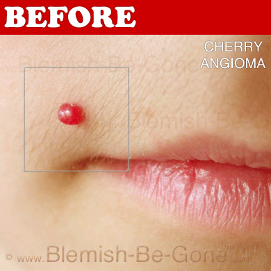
Cherry angiomas are made up of clusters of capillaries at the surface of the skin, forming a tiny round dome (” papule”), which may be flat topped. They range in color from bright red to purple. When they initially develop, they may be only a tenth of a millimeter in diameter and practically flat, appearing as small red dots. Nonetheless, they then typically increase to about one or two millimeters throughout, and often to a centimeter or even more in diameter. As they grow larger, they have a tendency to increase in thickness, and could take on the raised and rounded design of a dome. Numerous adjoining angiomas are stated to form a polypoid angioma. Since the blood vessels comprising an angioma are so close to the skin’s surface, cherry angiomas may bleed profusely if they are hurt.
One study located that the greater part of capillaries in cherry hemangiomas are fenestrated because of staining for carbonic anhydrase activity.
Click Here for Campbell de Morgan Spots removal.
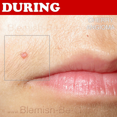
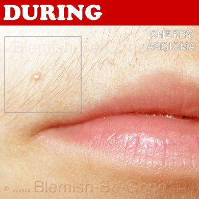
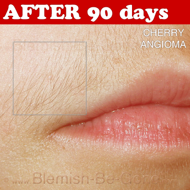
Campbell de Morgan Spots Pictures and Treatment
 |
 |
 |
 |
Amazing Natural Cure for Campbell de Morgan spots, Click Here.
Causes of Campbell de Morgan Spots
Cherry angiomas appear spontaneously in many people in middle age but can also, although less typical, arise in young people. They are able to likewise arise in an aggressive eruptive way in any age. The underlying root cause for the advancement of cherry angiomas is not recognized, a great deal because of a lack of interest in the subject matter. This is probably because they hardly ever are created by an inner malignancy.
The 1st research seeking to bring light to the molecular and genetic mechanisms behind cherry/senile hemangioma was recently published. The research located that the degree of MicroRNA 424 is substantially diminished in senile hemangiomas compared to normal skin generating increased protein expression of MEK1 and Cyclin E1. By hindering mir-424 in regular endothelial cells they may note the same increased protein expression of MEK1 and Cyclin E1 which, necessary for the advancement of senile hemangioma; led to cell expansion of the endothelial cells. They also located that targeting MEK1 and Cyclin E1 by having little interfering RNA lowered the amount of endothelial cells.
Chemicals and compounds that have been seen to cause cherry angiomas are mustard gas, 5 2-butoxyethanol, 9 bromides and cyclosporine. 11
A considerable increase in the density of mast cells has been viewed in cherry hemangiomas compared with ordinary skin.
Treat unwanted Campbell de Morgan Spots, Click Here.
Campbell de Morgan Spots Treatment
Removing unwanted facial irritants can be as easy as finding the right cream or lotion. However some issues require more complex solutions, like a professional microdermabrasion machine for example. Make sure you educate yourself on what your specific situation calls for.
On the rare occasions that they need removal, traditionally cryosurgery or electrosurgery have been made use of. More recently pulsed dye laser or Intense Pulsed Light (IPL) treatment has even been made use of.
Future treatment based on a locally acting inhibitor of MEK1 and Cyclin E1 may possibly be a selection. A natural MEK1 inhibitor is Myricetin
Prognosis
In most sufferers, the number and size of cherry angiomas boosts by having advancing age. They are simple, except in extremely rare cases that involve an abrupt look of several angiomas, which can be an indication of an establishing internal malignancy.
Epidemiology of Campbell de Morgan Spots
Cherry angiomas take place in all races, ethnic backgrounds, and both sexes.
For Campbell de Morgan Spots treatment and removal, Click Here.
Related Keywords: campbell de morgan spots, campbell de morgan spots removal, campbell de morgan spots treatment, campbell de morgan spots pictures, campbell de morgan spots photos, pictures of campbell de morgan spots, campbell de morgan spots picture